


Ultrasound of blood vessels
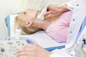
Vascular ultrasound uses sound waves to check and evaluate the body’s circulatory system, identify congestion and detect blood clots if any.
Nowadays, diseases related to veins and circulation become more and more complicated. If not examined, detected promptly will cause dangerous symptoms, difficult to treat. One of the methods that helps detect such abnormalities is Vascular ultrasound. Vascular ultrasound – a method of investigating and evaluating the circulatory system of the body.\
1. What is vascular ultrasound?
Vascular ultrasound is a method of using sound waves to check and evaluate the body’s circulatory system, identify congestion and detect the location of blood clots if any. Vascular ultrasound does not cause any adverse effects on the patient’s health.
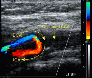
Ultrasound simulates images as well as all arterial and venous activities. When we have an ultrasound we can see if the structure, the size of the arteries, the veins, as well as the internal blood flow, are stable or anything abnormal.
2. Why is it necessary to vascular ultrasoundVascular ultrasound is one of the techniques that helps physicians check the flow of blood in tissues and organs in the body. From that, determine whether the blood flow is happening such as abnormal arteries, veins, plaque problems, blood clots …, clearly identify the narrow place to offer treatment options. , prevent fitting.
Not only assess the level and risk of cardiovascular diseases but also evaluate the effectiveness of previous treatment processes.
Specifically, this ultrasound is indicated for:
- Monitor blood flow to organs and tissues in the body.
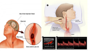
- Detect and locate abnormal obstructions and plaque or embolism and support the patient’s treatment plan.
- A blood clot is detected in the large veins of a leg or arm.
- Determine whether the patient is eligible for procedures such as coronary angioplasty.
- Assess the effectiveness of transplant surgery or blood vessel bypass.
- Determine whether the patient has an aneurysm.
- Determine the origin and severity of varicose veins.
 \
\
3. Where is a vascular ultrasound needed?
- Patients are prescribed ultrasound by a doctor when having the following signs:
- Deep vein thrombosis: This is a condition related to blood clots in the veins located deep inside the body. In people with deep venous valve failure, sedentary activities, the risk of embolism is very high.
- Arteriosclerosis: Arterial and narrowing arteries that supply blood to the legs and feet, affect the circulation of blood to the lower body.
- Superficial thrombophlebitis: A thrombophlebitis caused by blood clots that form in a superficial vein, just below the surface of the skin.
- Deep venous valve failure: This symptom reduces the quality of life of the patient. Early detection and prevention are most effective in the current medical context.
- There is a vascular tumor in the arm or leg position.
4. Procedure to perform vascular ultrasound
4.1 Preparation prior to vascular ultrasound
You should wear comfortable, loose-fitting clothes to the clinic for an angiogram. To perform an ultrasound, you may be asked to remove the jewelry and to change clothes from the clinic.
Ultrasound is difficult to perform if the patient is in constant motion. Therefore, hyperactive children or crying can make the ultrasound process difficult. To ensure smoothness, parents should instruct the baby beforehand. Bring books, toys to distract your baby’s interest and make time pass quickly.
4.2 The process of performing blood vessel ultrasound
In most cases of vascular ultrasound, you are placed on a flat surface. To improve the image quality, you may have to lie on your side or face down according to your doctor’s instructions. Next, apply water gel to the areas where ultrasound is performed to help the transducer make safe contact with the body and remove air pockets between the transducer and the skin that can block sound waves from entering the body.
Once done, change your clothes and wait for the doctor to review the ultrasound image. Ultrasound usually takes place within 15-30 minutes. However for complicated cases, this process can take longer.
The doctor will need to reduce the light in the ultrasound room to help make the observation of the image clearer,
4.3 What does vascular ultrasound need to do?
After the scan, if the doctor finds out the disease, you will be consulted enthusiastically about the next steps to take, if there is no problem, you will be allowed to join the doctor. daily activities.
5. Where is reputable blood vessel ultrasound in Tp. Ho Chi Minh
36 Ultrasound Clinic is one of the prestigious ultrasound addresses in Ho Chi Minh City. HCM. Since its establishment until now, Clinic has always strived in all medical activities to improve the quality of image diagnosis, healthcare for patients, with strengths such as:
5.1 Clinic equipment:
All modern machinery and equipment are 100% newly invested and imported from G7 countries with modern and accurate technology.
5.2. Team of good, experienced doctors:
Being diagnosed by leading experts, experienced professors and doctors, regularly updating knowledge, advanced healthcare methods and modern medical techniques
5.3 Public and transparent costs:
All expenses at 36 Ultrasound Clinic are always publicly and transparently posted, suitable for all patients.
If you have any questions, please contact our 36 Ultrasound Clinic hotline: 0917717498; email: phongkhamsieuam36@gmail.com or directly to the clinic at 36 Street 6, Khang An Resident Area, Phu Huu Ward, Thu Duc City, HCM City.