


Echocardiography
Echocardiography is one of the important methods used to check for abnormalities in the heart, in the diagnosis of heart conditions, showing the size, thickness, ability to pump blood and other activities of the heart. heart. The following article will help you learn more about this method.

1. Learn general about echocardiography
1.1 What does an echocardiogram show?
By ultrasound, the doctor can observe the heart’s structure and also check for abnormalities when the heart is functioning. Specifically, lets know:
-The way the heart works, contracts.
-Size and shape heart.
-The size and pumping motion of the heart walls.
-The pump of the heart.
-Does the heart valve work properly?
-Is the heart valve narrowed?
-Have blood regained through the heart valve (open valve).
-There’s a tumor, an inflammatory mass around the heart valve, heart contraction, no blood vessels.
Thus, with this information, the doctor will be able to diagnose problems encountered in the heart such as:
– Problems with large blood vessels entering and leaving the heart.
– Cardiac muscle problems, inner and outer cardiac membrane layers.
-The heart valve disease
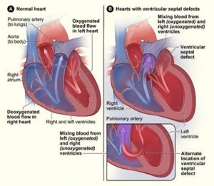
-The abnormal holes between the heart chambers
– Department of blood clots in the heart chamber.
1.2 When do you need an echocardiogram?
Your doctor will ask you to have an echocardiogram for a variety of reasons, for example when a heart is found to be abnormal through other tests, or if you have a heart condition, or a heart stethoscope.
In addition, if you have abnormal heart signs such as shortness of breath or chest pain, an echocardiogram is needed.
2. Procedure of echocardiography
2.1 Preparation before an echocardiogram
With conventional echocardiography, you do not need to prepare anything to eat, drink and take medicine as usual.
2.2 Procedure of echocardiography
Most echocardiography procedures take less than an hour, and the procedure may take longer or be shorter depending on your condition.
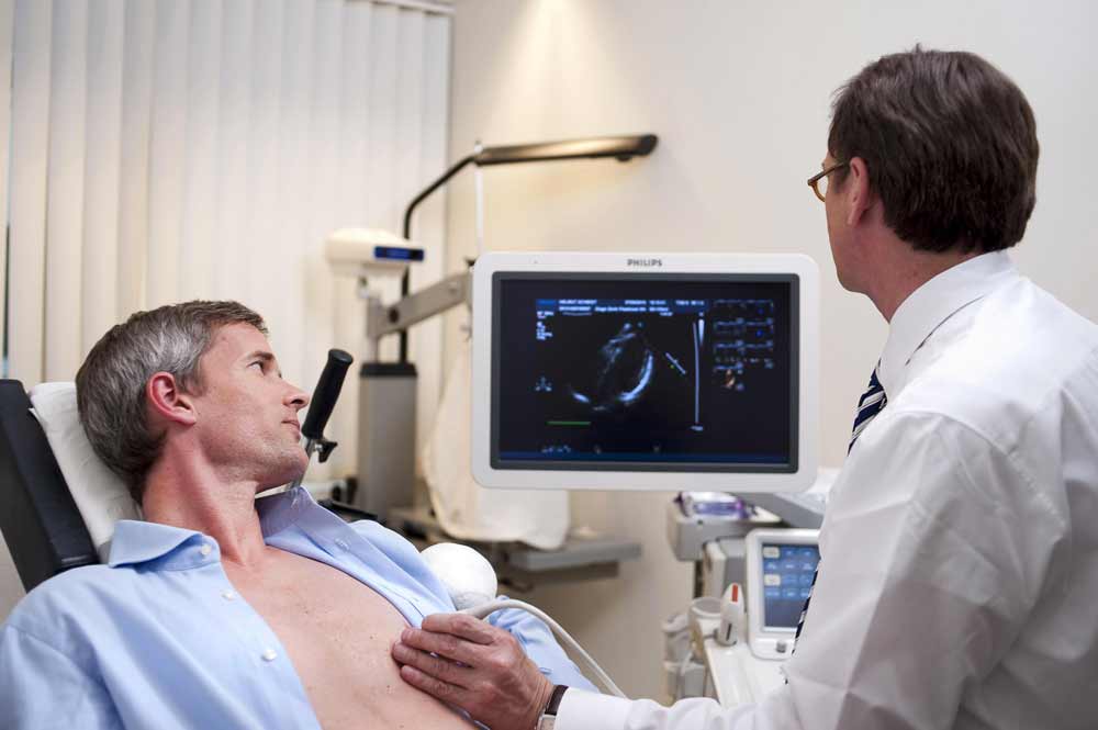
Perform an echocardiogram at the clinic. At this point, you are asked to lie on the bed, pulling the shirt from the waist up
Your doctor will need to reduce the light in the ultrasound room to help you see the image more clearly, then you will apply a special gel on your chest to increase the ability to conduct ultrasound.
The probe is moved back and forth on the chest to record echocardiogram images. You can hear píu píu, the sound of blood flowing in the heart that the ultrasound machine records.
2.3 What should I do after an echocardiogram?
After performing an echocardiogram, if the doctor finds out the disease you will be consulted enthusiastically about the next steps to take, if there is no problem, you will be allowed to join the doctor. daily activities.
3. Where echocardiography reputation in the city. Ho Chi Minh
36 Ultrasound Clinic is one of the prestigious ultrasound addresses in Ho Chi Minh City. HCM. Since its establishment until now, Clinic has always strived in all medical activities to improve the quality of image diagnosis, healthcare for patients, with strengths such as:
3.1 Clinic equipment:
All modern machinery and equipment are 100% new invested and imported from G7 countries with modern and accurate technology.
3.2. Team of good, experienced doctors:
Being diagnosed by leading experts, experienced professors and doctors, regularly updating knowledge, advanced healthcare methods and modern medical techniques
3.3 Public and transparent costs:
All expenses at 36 Ultrasound Clinic are always publicly and transparently posted, suitable for all patients.
If you have any questions, please contact our 36 Ultrasound Clinic hotline: 0917717498; email: phongkhamsieuam36@gmail.com or directly to the clinic at 36 Street 6, Khang An Resident Area, Phu Huu Ward, Thu Duc City, HCM City.