


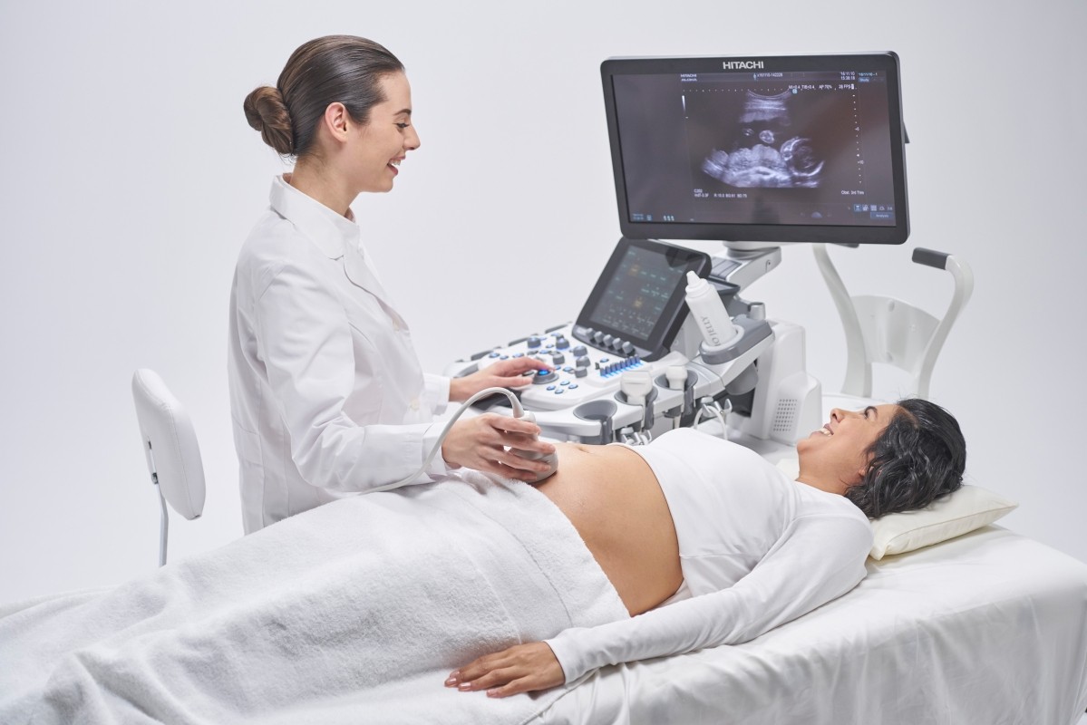
Fetal ultrasound
Fetal ultrasound is a non-invasive form of medical diagnostic test. By using sound waves, fetal ultrasound can show images of the baby in the abdomen. The ultrasound will transmit sound waves through the baby’s uterus and body, then through the computer reproduce into images that show the shape, position and movements of the baby. Not only the height and weight of the fetus, fetal ultrasound can also detect congenital malformations at certain stages of pregnancy. Women who are pregnant with diabetes, high blood pressure or who have special health problems may be ordered to have a higher pregnancy ultrasound.

1. Purpose of pregnancy ultrasound
– Check the position of the fetus (inside or outside the uterus)
– Calculate gestational age, weight and track fetal growth
– Check and detect fetal defects early (if any)
– Expected date of birth.
– Fetal monitoring for special tests such as laparoscopy, amniocentesis
2. When was the first ultrasound of pregnancy?
After detecting early signs of pregnancy such as missed periods, blood reports, 2-line pregnancy test …, you can go to the clinic to check if you are pregnant or not. This will be the first ultrasound scan to help the doctor identify how many weeks old the fetus is. Fetal ultrasound during this period also helps the doctor to determine whether the fetus has entered the uterus, avoiding ectopic pregnancy.

3. The important pregnancy ultrasound timeline
As recommended by the World Health Organization, fetal ultrasound is recommended to be conducted at least 3 times throughout the entire pregnancy. Inside:
– 1st fetal ultrasound should be conducted when the fetus reaches 11 to 13 weeks of age. First fetal ultrasound helps determine gestational age, prognosis of fetal development and expected relatively accurate delivery, Besides, the first fetal ultrasound is the only time to determine the nuchal translucency (a sign of DOWN syndrome screening). The results of the 1st fetal ultrasound help the doctor make an order to conduct a prenatal diagnostic test related to certain genetic defects / fetal chromosomes
– The second fetal ultrasound should be conducted at 20 to 24 weeks, at this stage, the morphological abnormalities of the fetus can be clearly identified. Therefore, in addition to evaluating the fetal morphological development, the second fetal ultrasound plays an important role to determine the risk of congenital fetal diseases as well as to evaluate fetal body abnormalities. Children such as malformation of internal organs, cleft palate, cleft lip … from which clinicians have specific advice on pregnancy care for pregnant women
– The 3rd fetal ultrasound should be conducted at 30 to 32 weeks, this is an important ultrasound to help the doctor predict the date of birth as well as predict the difficulties arising during the birth such as abnormal birth, weight During pregnancy, some of the possible deformities in the fetus, placenta, umbilical cord, amniotic fluid, etc. contribute to the detection of potential risks of obstetric complications.
4. Process of performing fetal ultrasound
4.1 Preparation before fetal ultrasound
– Drink plenty of fluids and stop urinating for 2 hours before the scan. As recommended by doctors, when the mother’s bladder is full of water, the ultrasound process will be easier. Images of babies in the womb are also shown more clearly.
– Wearing spacious and comfortable clothes
4.2 The process of performing fetal ultrasound
Most ultrasound procedures take less than an hour, and the procedure may take longer or be shorter depending on your condition.
Your doctor will need to reduce the light in the ultrasound room to help you see the image more clearly, then you will be applied a special gel to increase the ability to transmit ultrasound.
4.3 What should I do after an ultrasound scan?
After performing an echocardiogram, if the doctor finds suspicious signs you will be consulted enthusiastically about the next steps to take, if no problem occurs, you will be given a doctor’s permission to participate in daily activities.
5. Where pregnancy ultrasound reputation in the city. Ho Chi Minh
36 Ultrasound Clinic is one of the prestigious ultrasound addresses in Ho Chi Minh City. HCM. Since its establishment until now, Clinic has always strived in all medical activities to improve the quality of image diagnosis, healthcare for patients, with strengths such as:
5.1. Clinic equipment:
All modern machinery and equipment are 100% new invested and imported from G7 countries with modern and accurate technology.
5.2. Team of good, experienced doctors:
Being diagnosed by leading experts, experienced professors and doctors, regularly updating knowledge, advanced healthcare methods and modern medical techniques
5.3 Public and transparent costs:
All expenses at 36 Ultrasound Clinic are always publicly and transparently posted, suitable for all patients.
If you have any questions, please contact our 36 Ultrasound Clinic hotline: 0917717498; email: phongkhamsieuam36@gmail.com or directly to the clinic at 36 Street 6, Khang An Resident Area, Phu Huu Ward, Thu Duc City, HCM City.