


Breast ultrasound
Ultrasound is a commonly used diagnostic imaging technique, and breast ultrasound uses ultrasound to build and reconstruct images of the internal structure of the mammary glands and body. So far, this is considered a safe, simple, low-cost, non-toxic, non-invasive, fast, results-free, painless method that can be used for all ages, from children to old people. Therefore, breast ultrasound is widely used in the world and in Vietnam in examining, diagnosing and screening breast abnormalities, especially cancer.
1. Who needs breast ultrasound
Women over the age of 20 should regularly check breasts themselves. When performing regular self-examination, women will know the status of each breast so it is easy to distinguish between what is normal and what is abnormal, such as looking at a mirror to check the breast shape in a normal position, then switching positions Two hands raised and finally with hands on the hips, check the breast when changing positions. If there are detected lumps, swelling, pain, changes in skin color … abnormalities, you must go to the doctor for more specific examination and advice. For healthy women aged 35 and over, the doctor’s advice is to have regular breast exams including annual gynecological exams, preferably every 3 to 6 months, especially for women. have risk factors (People with a family history of breast cancer are also at higher risk. Women with early menstruation (under 12 years), late menopause (after 55 years); frequent use There is a high risk of taking birth control pills or taking estrogen instead, and women who do not have children or have their first child after age 30; or have a diet high in animal fat, drink alcohol, smoke. ; frequent exposure to radiation.) Particularly for breast ultrasound, should be checked at least every 6 months

2. Breast ultrasound can detect:
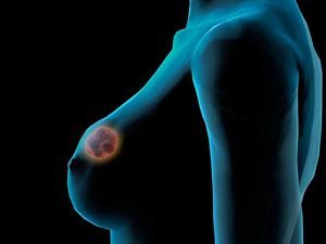
Breast ultrasound will help doctors detect abnormalities in an area on a breast that is suspected to have a tumor. This method will then be performed to continue the assessment and diagnosis to distinguish between a fluid-filled cyst and a dense breast. Besides, breast ultrasound can also help detect small lesions that are normally not visible or palpable. Also identify abnormalities in the mammary glands for pregnant women, when other imaging devices are not applicable.
Breast ultrasound helps assess breast masses in female patients under the age of 35, in which case mammography often does not show clear results. In 2003, recognizing that ultrasound was widely used in breast imaging, the American Photovoltaic Association (ACR) made the first definition of BI-RADS (Breast Imaging Reporting and Data System) in ultrasound. breasts, to standardize the visual interpretation of breast lesions. Today there are many BIRADS classifications, such as ACR 2013 classifications, Kim et al. 2008 classification, etc., based on 6 morphological characteristics of solid breast masses: shape, direction, shore, lesion boundaries, inner sound structure, characteristics of posterior sounds, with benign and malignant signs, from which conclusions are evaluated about the nature of the lesion.
Breast ultrasound can also help detect breast cancer growths in the breast. These cases are usually indicated biopsy through ultrasound guidance.
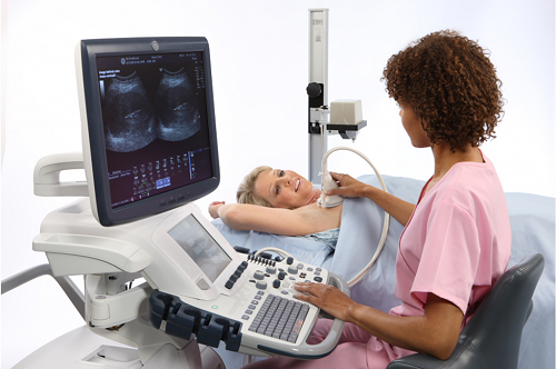
3. The process of performing breast ultrasound
3.1 Preparation before ultrasound transducer
With regular breast ultrasound, you don’t need any special preparation and can eat, drink and take your medicine as usual.
3.2 The process of performing breast ultrasound
Most ultrasound procedures take less than an hour, and the procedure may take longer or be shorter depending on your condition.
The doctor will need to reduce the light in the ultrasound room to help make the visual observations clearer,
3.3 What should I do after an ultrasound scan?
After the scan, if the doctor finds out the disease, you will be consulted enthusiastically about the next steps to take, if there is no problem, you will be allowed to join the doctor. daily activities.
4. Where breast ultrasound reputation in the city. Ho Chi Minh
36 Ultrasound Clinic is one of the prestigious ultrasound addresses in Ho Chi Minh City. HCM. Since its establishment until now, Clinic has always strived in all medical activities to improve the quality of image diagnosis, healthcare for patients, with strengths such as:
4. 1 Clinic equipment and facilities:
All modern machinery and equipment are 100% new invested and imported from G7 countries with modern and accurate technology. 4.2. Team of good, experienced doctors:
4.2 Team of good, experienced doctors:
Being diagnosed by leading experts, experienced professors and doctors, regularly updating knowledge, advanced healthcare methods and modern medical techniques
4.2 Public and transparent costs:
All expenses at 36 Ultrasound Clinic are always publicly and transparently posted, suitable for all patients.
If you have any questions, please contact our 36 Ultrasound Clinic hotline: 0917717498; email: phongkhamsieuam36@gmail.com or directly to the clinic at 36 Street 6, Khang An Resident Area, Phu Huu Ward, Thu Duc City, HCM City.